
Developed by a German chemist named Gustav Giemsa, the Giemsa stain is a type of Romanowsky stain. Originally intended for testing blood smears for malaria parasites, it is also used in histology to examine blood smears routinely. It is one of the most popular microscopic stains and thus its utility is well established in hematology for blood and bone marrow specimens, bacteriology, clinical cytology specimens, histological biopsies, and tumor samples.
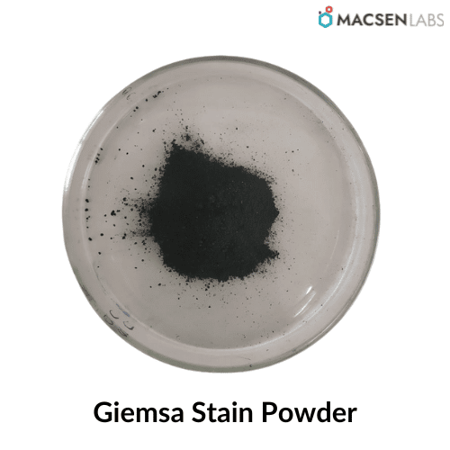
Table of Contents
Properties of Giemsa stain
| Property | Description |
|---|---|
| Product name | Giemsa Stain |
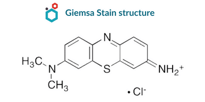
| IUPAC name | 7-imino-N,N-dimethylphenothiazin-3-amine;hydrochloride |
| Synonyms | Mixture of Azure II Eosinate & Methylene Blue; mancha de giemsa; tinción de giemsa; giemsa labe; tache de giemsa |
| Chemical Formula | C14H14ClN3S |
| CAS No. | 51811-82-6 |
| Molecular Weight | 291.8 g/mol |
| Appearance | Dark Green to black crystal or powder |
| Melting point | 300 °C (lit.) |
Composition
The basic constituents of Giemsa stain are the same; however, dilutions can be prepared based on their intended purpose.
| INGREDIENTS | gm/L |
|---|---|
| Giemsa Powder | 7.6 |
| Glycerol | 500ml |
| Methanol | 500ml |
Further, Giemsa stain is prepared with the composition of eosin and methylene blue–azure.
Principle (How Giemsa stain works)
The Giemsa stain is a differential stain that includes a combination of eosin dye, methylene blue, and azure in its composition. It binds specifically to the phosphate groups of DNA and does so in regions with a high concentration of the adenine–thymine interaction that is characteristic of DNA.
Both azure and eosin are types of acidic dye that can leave varying degrees of staining on the fundamental components of cells, such as the cytoplasm and granules. Methylene blue is the basic dye that is responsible for staining the acidic components of the cell, particularly the nucleus. In addition to its role as a stain for cells, methanol can also be used to “fix” an image. The cells are able to stick to the glass slide due to the fixative, preventing any additional changes in the cells from taking place.
Procedure
There are so many purposes for which specifically Giemsa stain is used.
For In-house preparation of stain
- Place 90 ml of buffered water into the tube.
- Filter the Giemsa stock solution through paper Whatman and transfer it to the container.
- Add the solution using a dry pipette.
- Prepare the Giemsa working solution before staining blood film and use it within 15 minutes of preparation.
For staining slides
Staining slides involves three methods and procedures explained below:
For Thin blood smears
Thin blood smears use 1:20 dilution and the procedure includes:
- Dip the film briefly in absolute methanol in a Coplin jar.
- Take out and let it dry.
- Then stain with diluted Giemsa stain in a Coplin jar.
- Briefly dip the slide in and out to wash it.
- Keep in a vertical position and air dry.
For Thick blood smears
- Dry the film for several hours and avoid by an incubator or by heat.
- Do not fix and stain with the diluted Giemsa stain.
- Then wash the film with water.
- Let it air dry and observe under the microscope using an oil immersion lens.
For Chlamydia trachomatis
The steps continue to be the same as for thin and thick smear but with the dilute stain of 1:40 dilution that was previously for 1:50 for thick and 1:20 for thin and leave the stain for 1-2 hours.
Observations
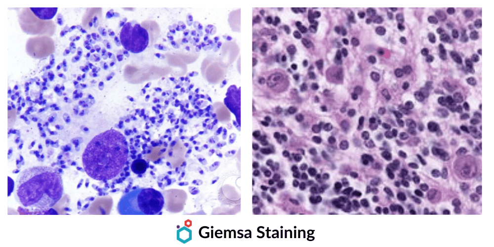
Under the microscope, this specific result comes out when bacteria, cell organelles, and parasites are distinguished on the basis of morphology and color.
CELL COMPONENTS- COLOR OBSERVED POST STAINING
- Red Blood Cells: Mauve-pink
- Neutrophils: Reddish purple
- Eosinophils: Purple nuclei & red to orange granules
- Basophils: Purple nuclei & blue coarse granules
- Lymphocytes: Dark blue nucleus
- Platelets: Violet color granules
- Nuclei of host cells: Dark purple
- Nuclei of WBCs: Dark purple
- The cytoplasm of host cells: Pale blue
- The cytoplasm of white cells: Pale blue or grey blue
- Melanin granules: Black green
- Bacteria: Pale or dark blue
- Borrelia spirochetes: Mauve-purple
- Malaria parasite: Red or pink nucleus and blue cytoplasm
- Monocytes: Pink Cytoplasm
Uses
Specifically, it binds to DNA regions with high adenine-thymine bonding levels and attaches to phosphate groups. Giemsa stain is used to create a karyogram or chromosome map by staining chromosomes in Giemsa banding, commonly called G-banding. A translocation or rearrangement can be detected by this method. The main use of Giemsa Stain is staining malarial parasites but apart from that, it has multiple uses and applications in Microbiology and pathology.
- It is used to obtain white blood cells.
- Platelets, RBCs, and WBCs are differentiated by this method with nuclear and cytoplasmic morphology.
- In Microbiology, giemsa stain is used for staining Chlamydia trachomatis, and Borrelia species.
- It is also used to identify chromosomal aberrations and in cytogenetics and also used for G-banding.
FAQs
Q. Which structures does Giemsa Stain identify?
Giemsa is used to identify the mast cells and stains the fungus Histoplasma, and Chlamydia bacteria.
Q. What is the difference between Giemsa stain and wright stain?
Giemsa stain is used to identify chromosome aberration by staining the chromosomes and wright stain is used to identify the different blood cell types.
Q. What is May Grunwald Giemsa stain and what are it’s uses?
May Grunwald-Giemsa or MCG stain is a type of Romanowsky stain used for staining blood, bone marrow smears, and clinical cytological specimens. This method is used for differential counting of blood cells and morphological inspection.
Q. Is Giemsa stain positive or negative?
The Giemsa stain is positive and is usually confirmed by the traditional staining method.
Q. Why Giemsa stain is used for malaria?
Giemsa Stain is used in malaria diagnosis. It is the recommended and most reliable procedure for staining thick and thin blood films from the blood sample of the patient, for precise identification of the causative malaria species.
Q. What is the difference between Leishman stain and Giemsa stain?
Leishman stain provides clear visualization of the nuclear chromatin pattern of cells and is used for staining blood and bone marrow whereas Giemsa stain is used for staining the blood cells of hematopoietic tissues and is performed on paraffin sections.
Q. What is the function of glycerol in Giemsa stain?
Giemsa stain is used in staining blood cells and bacteria that is improved by stabilizing the dye solution with glycerol and is allowed for staining of cells for microscopy purposes.
Resources
Biological Stains | Classification, Examples & Uses
Disclaimer-
The information provided here is based on general knowledge, articles, research publications etc. and we do not claim the authenticity of any of the information provided above. We do not claim or suggest/advise any medical, therapeutic, health or nutritional benefits of Giemsa Stain. We do not supply or promote our Giemsa Stain product for the applications which are covered by valid patents and which are not approved by the FDA.
Macsen Labs is a manufacturer and supplier of high-quality Giemsa Stain.